
| Synonym: | 4′,6-Diamidino-2-phenylindole, dihydrochloride |
| CAS #: | 28718-90-3 |
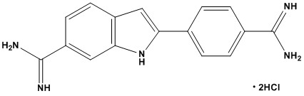
| Molecular Formula: | C16H15N5•2HCl |
| Molecular Weight: | 350.3 |
| DAPI, or 4′,6-diamidino-2-phenylindole, is a fluorescent stain that is a cornerstone of cell biology and microscopy. Its ability to specifically bind to DNA and emit a bright blue fluorescence has made it an indispensable tool for researchers. 1. Chemical Structure and Properties • Appearance: Typically a yellow-orange to dark brown powder or crystalline solid. • Solubility: Highly soluble in water (as the dihydrochloride salt), dimethyl sulfoxide (DMSO), and dimethylformamide (DMF). It is sparingly soluble in methanol and ethanol and insoluble in non-polar solvents. • Spectral Properties: Absorption Maximum (Excitation): ~358 nm (UV). Emission Maximum: ~461 nm (Blue/Cyan). Note: The binding mode affects its fluorescence. When bound to dsDNA, its fluorescence intensity enhances ~20-fold compared to its unbound state. Binding to RNA causes a redshift in excitation and emission. • Storage: Should be stored desiccated at -20°C, protected from light. Aqueous stock solutions are stable for months at 2-6°C in the dark but are susceptible to microbial growth; sterile filtration or addition of preservatives (e.g., sodium azide) is recommended for long-term storage. 2. Mechanism of Action and Specificity DAPI’s utility stems from its specific binding modes to nucleic acids: • Primary Mode: Minor Groove Binding to AT-Rich DNA DAPI binds preferentially to the minor groove of double-stranded DNA (dsDNA), with a strong affinity for clusters of adenine-thymine (A-T) base pairs. This binding results in a significant enhancement of its fluorescence quantum yield. • Secondary Mode: RNA Binding and Specificity Concerns DAPI can also bind to double-stranded RNA (dsRNA) and, to a lesser extent, single-stranded RNA, but with a different fluorescent signature (excitation ~400 nm, emission ~500 nm). This property is often considered an interference in DNA quantification but can be exploited for RNA staining under controlled conditions. • Non-Specific Binding: At higher concentrations, DAPI can stain other polyanionic structures, such as glycosaminoglycans in cartilage and some phospholipids, but this is not its primary use. 3. Primary Applications and Uses DAPI is a cornerstone technique in cell biology and molecular genetics. 3.1 Nuclear Staining and Cell Biology • Routine Nuclear Counterstain: Its most common application is as a counterstain in fluorescence microscopy (immunofluorescence, FISH, tissue sections) to visualize the nucleus and determine cellular location. It clearly defines nuclear morphology (condensed in apoptosis, enlarged in mitosis). • Apoptosis Assays: DAPI can be used to identify apoptotic cells by revealing nuclear condensation and fragmentation. 3.2 Cytogenetics and Karyotyping • Chromosome Banding (DAPI-Banding): Due to its AT-selectivity, DAPI produces characteristic banding patterns on metaphase chromosomes (Q-banding-like patterns), which are useful for karyotyping and identifying chromosomal aberrations. 3.3 Flow Cytometry • DNA Content and Cell Cycle Analysis: DAPI is a popular stoichiometric DNA stain for flow cytometry. It binds in proportion to DNA content, allowing for the quantification of DNA and subsequent analysis of cell cycle phases (G0/G1, S, G2/M) and the detection of aneuploidy. 3.4 Microbiology • Bacterial and Fungal Enumeration: DAPI is used to stain and count microbial cells in environmental samples, clinical specimens, and food samples. 3.5 DNA Quantification (Historical/Limited): While largely superseded by more specific dyes like PicoGreen for solution-based assays, DAPI can be used for fluorometric DNA quantification, particularly for AT-rich DNA. 4. Protocols and Handling • Working Solution: A common working concentration for nuclear staining is 1 – 5 µg/mL in PBS or your chosen buffer. A 5-10 mg/mL stock solution in water or buffer is standard. • Staining Procedure: Incubation times are typically short (5-30 minutes) at room temperature. For fixed cells, a simple rinse after staining is sufficient. For live cells, the stain is often added directly to the media, but note that DAPI is toxic. • Compatibility: DAPI is compatible with a wide range of other fluorophores (e.g., FITC, TRITC, Alexa Fluor 488, 555, 594) due to its blue emission. It is a standard part of DAPI/FITC/TRITC filter sets on microscopes. • Quenching: Fluorescence can be quenched by iodide ions and other heavy atoms. 5. Advantages and Limitations 5.1 Advantages: • High Specificity for DNA: Excellent for distinguishing DNA from RNA with proper filter sets. • Bright Signal: Large fluorescence enhancement upon DNA binding. • Cell Permeability: Can stain live and fixed cells, though it is toxic. • Photostability: Relatively photostable compared to some other DNA stains like Hoechst 33342. • Cost-Effective: Inexpensive and widely available. 5.2 Limitations: • UV Excitation: Requires a UV-capable microscope and mercury/xenon arc lamp or UV laser, which can be more damaging to cells and tissues. • Toxicity: Not suitable for long-term live-cell imaging due to its DNA-binding properties, which can be mutagenic and interfere with biological processes. • RNA Binding: Can cause background fluorescence if not properly controlled. • AT-Bias: Not a truly neutral DNA stain; quantification can be skewed for samples with varying AT/GC content. 6. Comparative Notes & Alternatives • Hoechst 33342/33258: Also minor-groove AT-preferring; better live-cell permeability than DAPI; excite at 350–405 nm; blue emission. • Propidium iodide (PI), 7-AAD, SYTOX dyes: Membrane-impermeant, red-emitting viability/DNA dyes for 488/561-nm systems (no UV needed). • DRAQ5/DRAQ7/TO-PRO-3: Far-red DNA stains compatible with 633–647-nm lasers; excellent for multiplexing with GFP/YFP/RFP. DAPI (CAS 28718-90-3) remains one of the most fundamental and indispensable tools in the cell biologist’s arsenal. Its exceptional properties as a minor-groove binding, AT-selective DNA stain make it the gold standard for nuclear counterstaining, cytogenetic banding, and DNA content analysis. While its requirement for UV excitation and inherent mutagenicity are limitations, its brightness, specificity, and cost-effectiveness ensure its continued widespread use. For any researcher working with fluorescent microscopy or flow cytometry, DAPI is a critical reagent for revealing the fundamental architecture of the cell—its nucleus. References: 1. Overview of Fluorescent Dyes 2. DAPI 3. DAPI Dye Profile 4. DAPI: a DNA-specific fluorescent probe |
|
DAPI
For Research & Development use only. Not for testing and/or use on humans.

